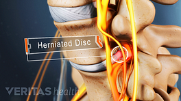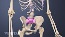
A thoracic herniated disc occurs when a spinal disc in the mid-back ruptures.
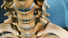
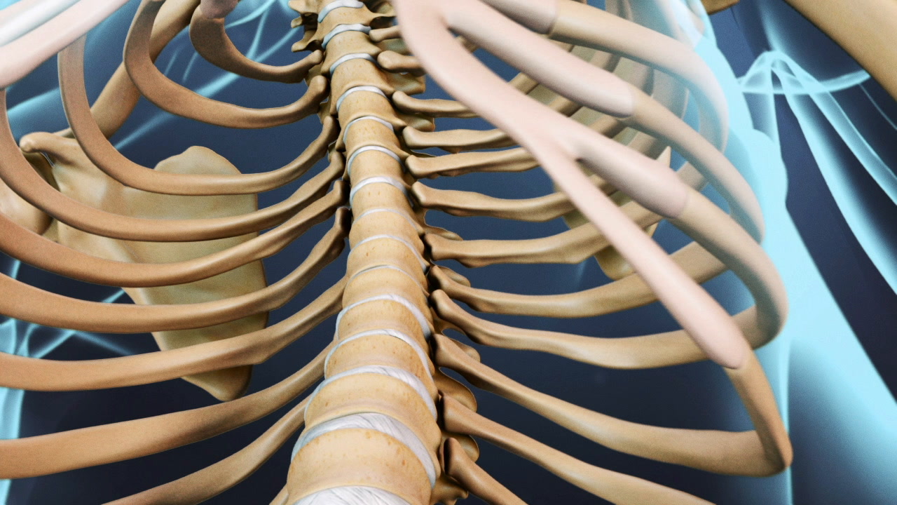
In the thoracic spine, or upper back, there are eleven spongy discs positioned between the bony vertebrae.
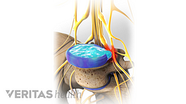
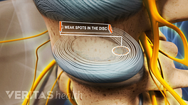
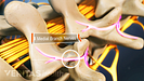
Each healthy disc in the spine contains a jelly-like inner core, called the nucleus pulposus, and a tough outer coating, called the annulus fibrosus.
In the center of the thoracic spine, the spinal cord travels through the spinal canal, and at each vertebral level a nerve root exits the canal.
When a thoracic disc herniates, the inner nucleus pulposus leaks out, which can impinge upon or cause inflammation near one of these exiting nerve roots.
When this happens, called a lateral herniation, it can cause radiating nerve pain along the path of the affected nerve, such as into the chest or abdomen.
If a thoracic disc herniates back into the spinal cord area, it may cause numbness below the level of cord compression, as well as difficulty with walking or balance, or possible loss of bowel and bladder control—a condition called myelopathy.
If a thoracic disc herniates both back into the spinal cord and to the side, called a centro-lateral herniation, symptoms may include a combination of radiating pain and myelopathy.
Thoracic disc herniations are rare. When they do occur, they tend to occur below the eighth thoracic vertebra, or T8 level, and the lowest thoracic segment is the most common.
Most thoracic herniated discs heal on their own or with nonsurgical treatment, but in some cases of myelopathy or intolerable pain, surgery may be recommended.