Percutaneous vertebroplasty is a minimally invasive procedure used to treat spinal compression fractures.
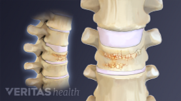
Compression fractures occur in spinal vertebrae that have been weakened by osteoporosis or other conditions that weaken the bones, such as cancer.
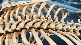
Compression fractures typically occur in the thoracic region of the spine, which includes the T1 through T12 vertebrae, but may also occur in the lumbar spine, or L1 through L5.
The goals of vertebroplasty are to reduce pain while also stabilizing and strengthening the fractured vertebra.
In the procedure, the patient lies face down on the operating table. The surgeon may reset the fractured vertebra by externally manipulating the patient's body position before surgery begins.
The surgeon punctures the skin over the affected vertebra with a biopsy needle.
Using X-ray guidance, the needle is inserted into the vertebra through the pedicle, or sometimes slightly above it. Using either a rotary motion or a tapping mallet, the surgeon pushes the needle into the bone at an angle until it reaches deep into the vertebral body.
Low-viscosity bone cement is injected into the vertebra, which then spreads throughout the weakened portion of the bone. Depending on how rapidly the cement spreads, a second injection may be necessary.
The cement hardens quickly, creating an internal cast inside of the fractured vertebra.
The patient may turn over and lie flat on his or her back while the cement hardens, which usually takes about 5 minutes. The entire process may be then repeated on the other side of the vertebral body.
The surgeon then bandages the skin puncture. Most patients can go home the same day as the procedure.
In This Article:
- Vertebroplasty After a Painful Spine Fracture
- Vertebroplasty Procedure
- Vertebroplasty Risks and Potential Complications
- Vertebroplasty Procedure Video