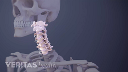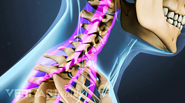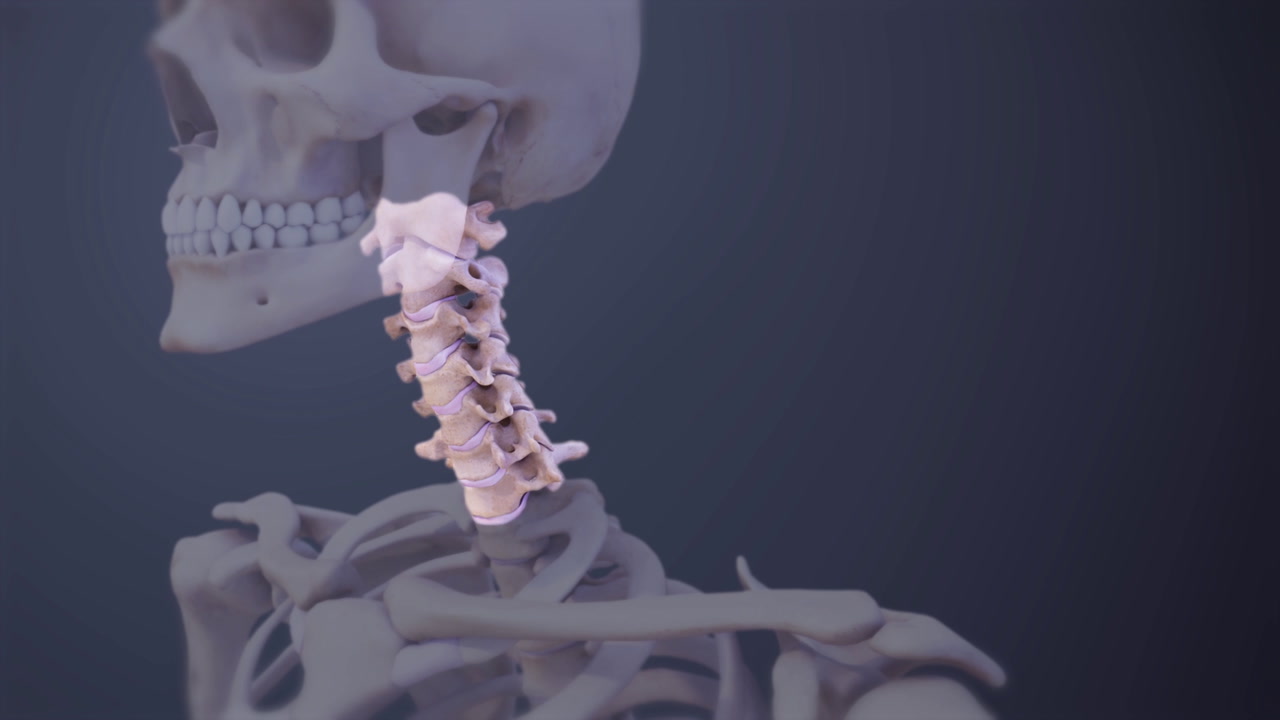
The neck has 7 cervical vertebrae, labeled C1 through C7. They form a natural inward curvature, sometimes called a lordotic curve. The upper cervical vertebrae are smaller and more mobile, while the lower ones are bigger to handle heavier loads from the neck and head.
C3 through C6 are considered typical vertebrae because they share the same basic features with most of the spine’s vertebrae. 1 Darby SA. General anatomy of the spinal cord. In: Cramer GC, Darby SA, Clinical Anatomy of the Spine, Spinal Cord, and Ans, 3rd Edition. St. Louis, MO: Elsevier; 2014: 122.
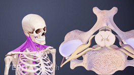
The vertebral body is the thick, oval segment of bone at the front of the vertebra and carries most of the load.
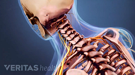
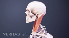
At the back, the vertebral arch consists of 2 pedicles and 2 laminae. The spinous process is a bony protrusion where muscles and ligaments attach. There is also a transverse process on each side. The spinal cord is protected within the vertebral foramen between the vertebral arch and vertebral body. If this foramen becomes narrowed, such as from spinal degeneration, it can cause central stenosis and compress the spinal cord.
A pair of facet joints connect adjacent vertebrae in the back. These joints are lined with smooth cartilage to enable limited movements. Facet joint osteoarthritis is a common cause of chronic neck pain.
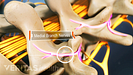
C3 through C7 are the only vertebrae to have uncovertebral joints, which help the neck’s forward and backward movements while limiting side bending. The uncovertebral joints are also a common location for bone spurs to develop as the spine ages and cause foraminal stenosis, which can compress a nearby spinal nerve.
C1 and C2 are considered atypical vertebrae.
The C1 vertebra, called the atlas, is shaped like a ring. It connects to the occipital bone above to support the skull and form the atlanto-occipital joint. Much of the head’s forward and backward motion occurs here. 1 Darby SA. General anatomy of the spinal cord. In: Cramer GC, Darby SA, Clinical Anatomy of the Spine, Spinal Cord, and Ans, 3rd Edition. St. Louis, MO: Elsevier; 2014: 122.
The C2 vertebra, called the axis, has a large bony protrusion known as the dens. The dens fits into the ring-shaped atlas above it, forming the atlantoaxial joint. Much of the head’s rotational motion occurs at this joint. 1 Darby SA. General anatomy of the spinal cord. In: Cramer GC, Darby SA, Clinical Anatomy of the Spine, Spinal Cord, and Ans, 3rd Edition. St. Louis, MO: Elsevier; 2014: 122. , 2 Heinking KP, Kappler RE. Cervical region. In: Chila A, ed. Foundations of Osteopathic Medicine. Philadelphia, PA: Lippincott, Williams & Wilkens. 2010: 513-527.
C7 is similar to the typical vertebrae, but it is commonly considered a unique vertebra. 1 Darby SA. General anatomy of the spinal cord. In: Cramer GC, Darby SA, Clinical Anatomy of the Spine, Spinal Cord, and Ans, 3rd Edition. St. Louis, MO: Elsevier; 2014: 122. One of C7’s distinguishing features is that its spinous process is longer so more muscles attach to it. 1 Darby SA. General anatomy of the spinal cord. In: Cramer GC, Darby SA, Clinical Anatomy of the Spine, Spinal Cord, and Ans, 3rd Edition. St. Louis, MO: Elsevier; 2014: 122. C7 is also the largest cervical vertebra and forms the base of the cervical spine. It connects with the thoracic spine below to form the cervicothoracic junction.
In This Article:
- 1 Darby SA. General anatomy of the spinal cord. In: Cramer GC, Darby SA, Clinical Anatomy of the Spine, Spinal Cord, and Ans, 3rd Edition. St. Louis, MO: Elsevier; 2014: 122.
- 2 Heinking KP, Kappler RE. Cervical region. In: Chila A, ed. Foundations of Osteopathic Medicine. Philadelphia, PA: Lippincott, Williams & Wilkens. 2010: 513-527.