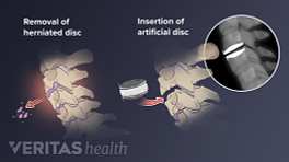
Cervical disc replacement surgery is designed to replace a damaged spinal disc in the neck with an artificial disc implant.
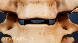
Damaged cervical discs are most likely to occur at the C4-C5, C5-C6, or C6-C7 levels.
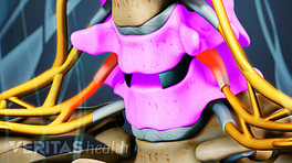
The damaged disc may be due to conditions such as a cervical disc herniation, and/or osteophytes, also called bone spurs, encroaching upon the nerve root.
During cervical disc replacement surgery, a one-to-two-inch incision is made in the front of the neck.
The thin platysma muscle that lies just underneath the skin is cut and moved aside, and the pre-vertebral fascia, a thin layer of tissue that surrounds the spine, is removed to expose the spinal discs.
An incision is made in the outer coating of the disc, called the annulus fibrosus, and the soft inner core of the disc, called the nucleus pulposus, is removed. Most of the damaged disc is extracted, but a small portion may be left intact.
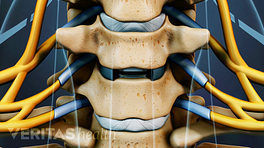
The disc space between the vertebrae is then restored to its normal height.
This restoration of disc space height aids in the decompression of surrounding nerve roots and makes room for the new artificial disc.
Using X-ray guidance, the surgeon inserts the artificial disc into the space previously occupied by the natural disc.
There are many different cervical artificial disc designs and materials commercially available.
Patients can usually leave the hospital after one to two days, with minimal activity restrictions.